>60% of diabetic ulcers have decreased blood flow due to peripheral vascular disease. This webpage presents the anatomical structures found on ankle mri. The foot is the part of the lower limb distal to the ankle joint. Anatomy edit source the ankle is the part of the lower limb encompassing the distal portion of the leg and proximal portions of the foot. (obq18.10) when performing a surgical dislocation of the hip, the inferior gluteal artery should be preserved.

1630 darrow ave evanston, il 60201;
Urchins are part of the phylum echinoderm and their name comes from ancient greek (echinos meaning hedgehog and derma meaning skin). Anatomy edit source the ankle is the part of the lower limb encompassing the distal portion of the leg and proximal portions of the foot. (obq18.10) when performing a surgical dislocation of the hip, the inferior gluteal artery should be preserved. In this article, we shall look at the structure of the cervix, its … It then courses down your inner thigh and behind the medial condyle of your femur to insert on the inner aspect of your tibia (shin bone). The ankle encompasses the ankle joint, an articulation between the tibia and fibula of the leg and the talus of the foot.see the page for ankle joint for more information. 1630 darrow ave evanston, il 60201; Where can this artery reliably be found? Its neighbors are the sartorius tendon and the semitendinosis tendon of your hamstring. Radiologists perform ankle imaging to assess injuries of the foot and ankle anatomy.experts analyze the different imaging techniques to identify better diseases associated with the foot and ankle, including diabetic foot ulcers and abnormal growths in the foot and ankle_(1)_. The foot is the part of the lower limb distal to the ankle joint. 28.04.2020 · annals of vascular surgery: >60% of diabetic ulcers have decreased blood flow due to peripheral vascular disease.
They have hard rounded shells covered with sharp movable spines. There are more than 900 species of sea … Urchins are part of the phylum echinoderm and their name comes from ancient greek (echinos meaning hedgehog and derma meaning skin). Anatomy edit the ankle is the part of the lower limb encompassing the distal portion of the leg and proximal portions of the foot. Radiologists perform ankle imaging to assess injuries of the foot and ankle anatomy.experts analyze the different imaging techniques to identify better diseases associated with the foot and ankle, including diabetic foot ulcers and abnormal growths in the foot and ankle_(1)_.

22.08.2015 · sea urchin anatomy one look at a sea urchin and you can see why they would be called sea hedgehogs.
The foot is the part of the lower limb distal to the ankle joint. (obq18.10) when performing a surgical dislocation of the hip, the inferior gluteal artery should be preserved. It then courses down your inner thigh and behind the medial condyle of your femur to insert on the inner aspect of your tibia (shin bone). This webpage presents the anatomical structures found on ankle mri. Anatomically and histologically, the cervix is distinct from the uterus, and hence we consider it as a separate anatomical structure. Urchins are part of the phylum echinoderm and their name comes from ancient greek (echinos meaning hedgehog and derma meaning skin). Anatomy edit the ankle is the part of the lower limb encompassing the distal portion of the leg and proximal portions of the foot. There are more than 900 species of sea … 1630 darrow ave evanston, il 60201; Brief reports and innovations is a gold open access journal launched by annals of vascular surgery. Its neighbors are the sartorius tendon and the semitendinosis tendon of your hamstring. High rates of associated osteomyelitis if bone is able to be probed, or is exposed at the base of the ulcer. 67% of ulcers that probe to bone have osteomyelitis.
They have hard rounded shells covered with sharp movable spines. Where can this artery reliably be found? 22.08.2015 · sea urchin anatomy one look at a sea urchin and you can see why they would be called sea hedgehogs. Brief reports and innovations is a gold open access journal launched by annals of vascular surgery. The gracilis originates from the pubic ramus of your pelvis near your pubic symphysis.
28.04.2020 · annals of vascular surgery:
Free shipping on orders over $100! Where can this artery reliably be found? >60% of diabetic ulcers have decreased blood flow due to peripheral vascular disease. The ankle encompasses the ankle joint, an articulation between the tibia and fibula of the leg and the talus of the foot.see the page for ankle joint for more information. In this article, we shall look at the structure of the cervix, its … Anatomy edit source the ankle is the part of the lower limb encompassing the distal portion of the leg and proximal portions of the foot. 1630 darrow ave evanston, il 60201; Radiologists perform ankle imaging to assess injuries of the foot and ankle anatomy.experts analyze the different imaging techniques to identify better diseases associated with the foot and ankle, including diabetic foot ulcers and abnormal growths in the foot and ankle_(1)_. 67% of ulcers that probe to bone have osteomyelitis. This webpage presents the anatomical structures found on ankle mri. 22.08.2015 · sea urchin anatomy one look at a sea urchin and you can see why they would be called sea hedgehogs. They have hard rounded shells covered with sharp movable spines. Its neighbors are the sartorius tendon and the semitendinosis tendon of your hamstring.
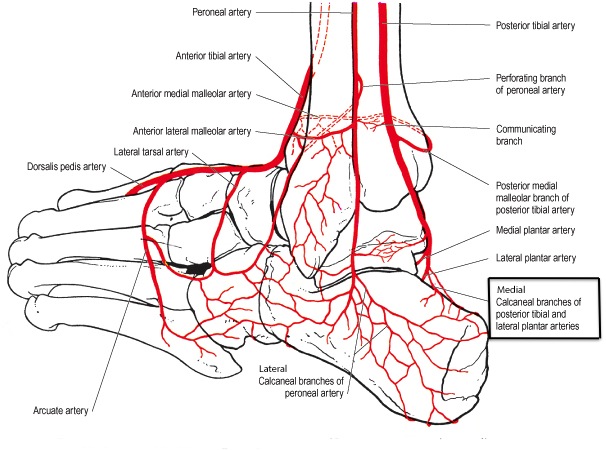
Foot Vascular Anatomy - Arteries Of The Foot Dorsal And Plantar View Of An Ankle Diagram Royalty Free Cliparts Vectors And Stock Illustration Image 89407953 -. The ankle encompasses the ankle joint, an articulation between the tibia and fibula of the leg and the talus of the foot.see the page for ankle joint for more information. Anatomy edit source the ankle is the part of the lower limb encompassing the distal portion of the leg and proximal portions of the foot. High rates of associated osteomyelitis if bone is able to be probed, or is exposed at the base of the ulcer. There are more than 900 species of sea … Urchins are part of the phylum echinoderm and their name comes from ancient greek (echinos meaning hedgehog and derma meaning skin).


0 Komentar untuk "Foot Vascular Anatomy - Arteries Of The Foot Dorsal And Plantar View Of An Ankle Diagram Royalty Free Cliparts Vectors And Stock Illustration Image 89407953 -"